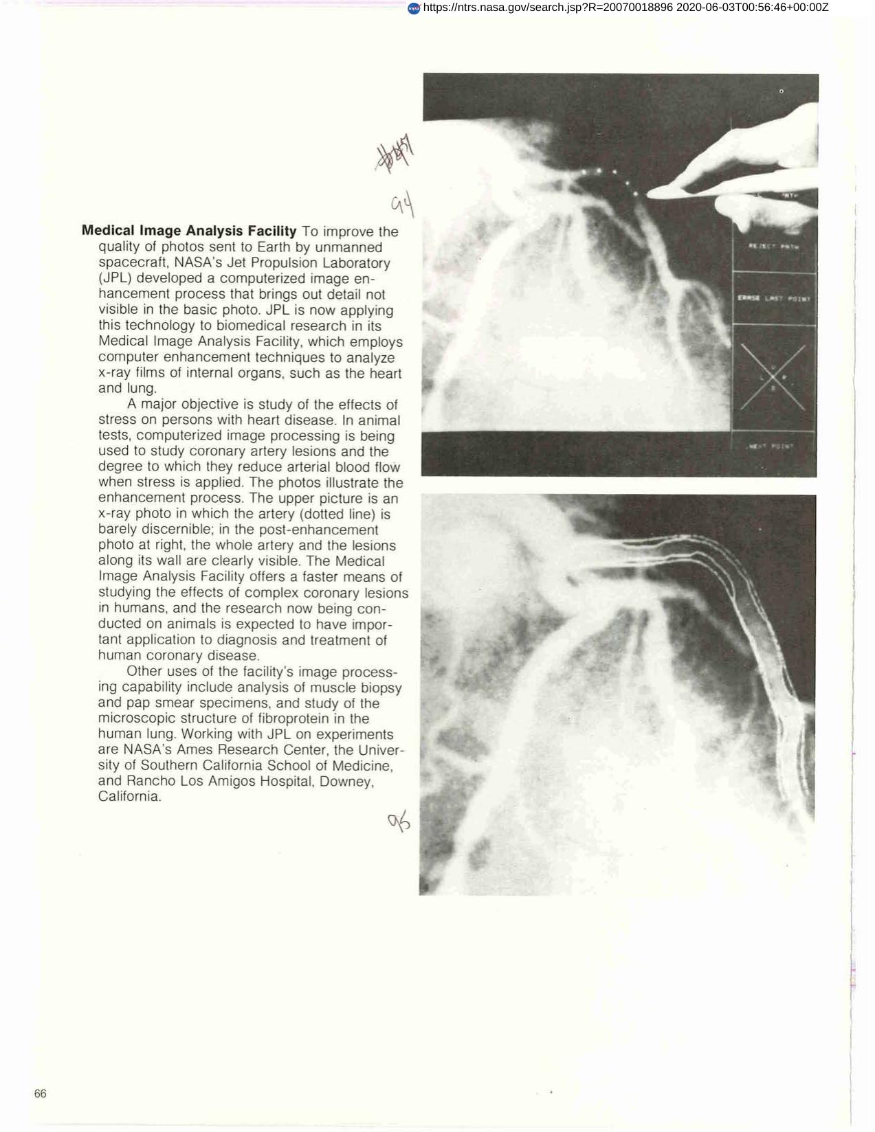

Medical Image Analysis Facility
Jet Propulsion Laboratory developed a computerized image enhancement process that brings out detail not visible in basic photos. This technology is now applied to biomedical research in JPL's Medical Image Analysis Facility which employs computer enhancement techniques to analyze x-ray films of internal organs such as the heart and lungs. Major objective is to study effects of stress on persons with heart disease. In annual tests, computerized image processing is being used to study coronary artery lesions, the degree to which they reduce arterial lesions, and the degree to which they reduce arterial blood flow when stress is applied.
Full article: http://hdl.handle.net/hdl:2060/20070018896
Medical Image Analysis Facility
Medical Image Analysis Facility